

A revolutionary Tomographic Microscope to look instantly inside living cells in 3D.
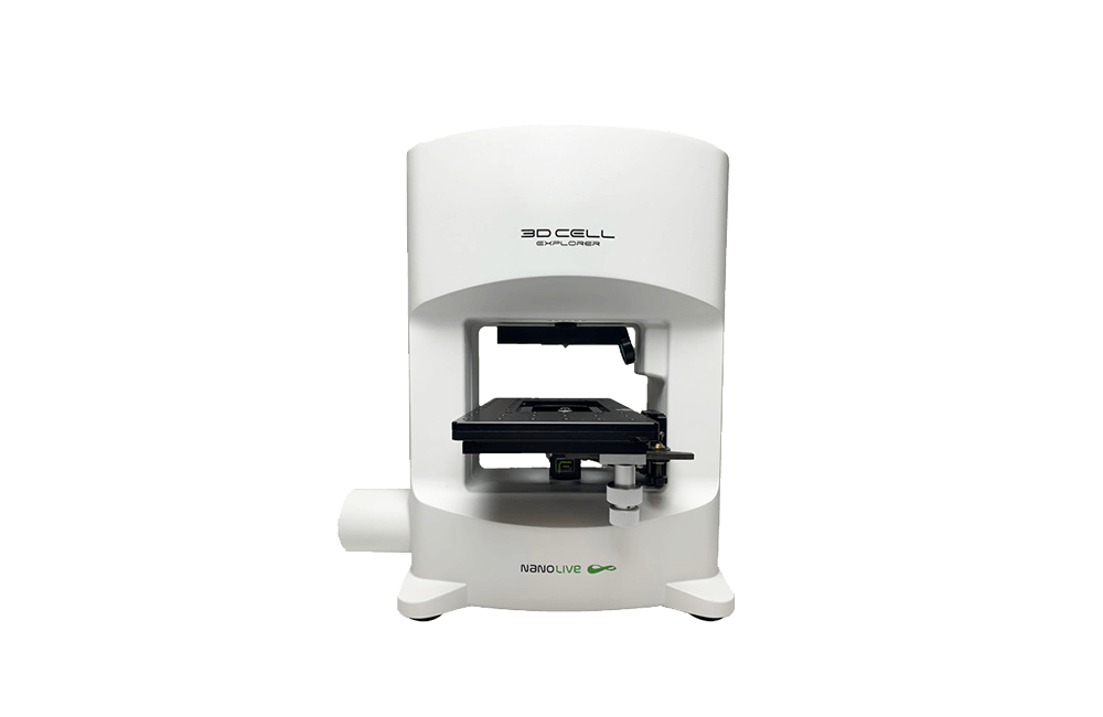
A live cell imaging microscope that can…
Analyze the cell’s inner structure and sub-structure in a non-invasive way: Explore and measure cell organelles with unprecedented detail and resolution, marker-free and preparation-free based on their own physical density.
Study cell life cycle processes of growth, division & death in 3D and 4D: Monitor all cell compartments and their kinetics and dynamics in real-time at every second without interfering with natural functioning.
Keep your sample healthy as long as you need: Thanks to a dedicated top-stage incubator you can keep your cells in a controlled environment and physiological conditions while imaging them.
NON-INVASIVE 3D CHARACTERIZATION
Live cell imaging in physiological conditions without any bleaching or phototoxicity
LABEL-FREE 4D CONTINUOUS OBSERVATION
Measurement of cell processes from seconds to weeks
MULTIPLEXING
High resolution and high sensitivity characterization of multiple cell organelles based on their refractive index
| Resolution | Δx,y = 200nm; Δz = 400nm |
| Field of View | 85 x 85 x 30 μm |
| Tomography frame rate | 0.5 fps 3D image rate with full self-adjustement |
| Microscope Objective | Air with 60x magnification |
| Illumination Source | Class 1 laser low power (λ=520 nm, sample exposure 0.2 mW/mm2) |
| Accessible Sample Stage | 60 mm of free access to the sample stage for sample manipulation |
| Dimensions | 38 cm x 17 cm x 45 cm |